Is the Brain ‘Two Organs’? What the Stanford Study Actually Found
Smartor 편집팀
September 18, 2026

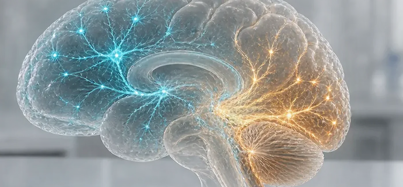
The claim that the human brain is “really two organs” sounds dramatic, but it does not mean that people have two separate brains or two independent minds. The central finding of a study published in Nature Neuroscience on September 18, 2026, is developmental: the forebrain and midbrain may arise from one neural ectoderm progenitor lineage, while the hindbrain arises from another lineage that appears alongside it early in embryonic development.
The research team traced cell lineages in mouse embryos and used human pluripotent stem cells to test how the two developmental routes behave. The results support a model in which anterior and posterior neural ectoderm progenitors appear in parallel, have different chromatin states, and are already restricted toward different brain-region fates. The researchers also used that insight to generate a type of human hindbrain motor neuron that had been difficult to produce reliably in the laboratory.
This is not a new clinical guideline. It does not mean that healthy people need brain scans, genetic tests, or different medication. It also does not show that amyotrophic lateral sclerosis (ALS) or spinal muscular atrophy (SMA) has been cured. Its immediate value is in basic and translational research: a more accurate way to make disease-relevant hindbrain cells could improve laboratory models used to study how certain disorders develop and how experimental treatments affect those cells.
This article provides general educational information and is not a diagnosis or treatment recommendation. New facial drooping, weakness on one side, trouble speaking, sudden severe headache, altered consciousness, breathing difficulty, or trouble swallowing may require urgent medical evaluation regardless of any research headline.
The adult brain is often divided into the forebrain, midbrain, and hindbrain. The forebrain includes structures involved in language, planning, memory, sensory integration, and conscious thought. The midbrain contributes to movement, alertness, and visual and auditory responses. The hindbrain includes structures such as the pons, medulla, and cerebellum and participates in breathing, heart-rate regulation, sleep, balance, swallowing, and the control of facial and throat muscles.
Those familiar divisions describe anatomy and function. The new paper asks a much earlier developmental question: do all brain regions descend from one original neural ectoderm progenitor population, or do multiple progenitor populations begin with different regional limits? A common model held that an early neural population later divides into the forebrain, midbrain, and hindbrain. The researchers instead found evidence supporting two progenitor lineages that emerge at roughly the same stage during gastrulation.
In mouse embryos, the team used lineage-tracing approaches to follow what early cells became. Cells expressing the gene Otx2 were associated with anterior neural ectoderm destined for the forebrain and midbrain. Cells expressing Gbx2 were associated with posterior neural ectoderm committed to the hindbrain. According to the paper and Stanford Medicine’s account, the populations were mutually exclusive at the stages examined rather than representing one population that simply switched identity later.
The researchers also examined chromatin, the packaged form of DNA and its associated proteins. Chromatin accessibility helps determine which genes are available for a cell to use. The anterior and posterior progenitors had different chromatin landscapes that anticipated their later forebrain/midbrain or hindbrain identities. In practical terms, the two groups were not merely in different places. Their molecular states had already placed them on different developmental tracks.
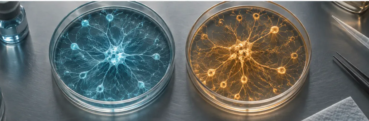
The team then differentiated human pluripotent stem cells into cells resembling anterior or posterior neural ectoderm. Those cells showed restricted tendencies toward forebrain/midbrain or hindbrain fates. By beginning with the posterior route, the researchers produced hindbrain rhombomere 5/6-specific motor neurons. These laboratory-grown cells generated action potentials and expressed proteins associated with hindbrain segments that control facial and swallowing muscles. That result matters because researchers had struggled to generate some authentic hindbrain neuron types using protocols that may have started with the wrong progenitor identity.
The paper also proposes an evolutionary interpretation. Comparisons involving chickens, zebrafish, and acorn worms suggest that dual neural origins may have been conserved for roughly 550 million years. The Stanford researchers describe the modern brain as two ancient neural systems brought together spatially through evolution. That is a scientific model supported by the reported comparisons, not proof that every nervous system in every species follows an identical program. Direct studies across additional species and developmental stages will be needed.
The phrase “two organs” emphasizes developmental lineage and evolutionary history. In an adult, the forebrain, midbrain, and hindbrain are tightly connected through neural circuits, blood vessels, glial support, and continuous anatomy. They work as an integrated nervous system. The study did not investigate the left and right hemispheres, and it does not validate popular claims that people have a “logical brain” and an “emotional brain.” It also does not propose a new diagnosis based on having two separate organs inside the skull.
A more accurate plain-language translation is: different major regions of the brain may begin from two distinct progenitor lineages early in development. That wording preserves what is new without inviting unsupported claims about personality, consciousness, or hidden brain capacity.
The most immediate effect is in the laboratory. Scientists cannot safely and repeatedly collect living human brainstem tissue for routine experiments. If patient-derived or genetically engineered pluripotent stem cells can be turned into the relevant hindbrain motor neurons, researchers can watch when disease-related changes begin, compare affected and unaffected cells, and test how experimental compounds alter cell function.
Producing a more faithful cell type can improve the relevance of an experiment, but a dish of neurons cannot reproduce an entire patient. ALS and SMA involve interactions among neurons, muscles, glia, immune cells, blood vessels, genetics, and the surrounding environment. The new method may offer a sharper view of one important part of the disease process without modeling every component of the human body.
For patients and families, this is encouraging basic research, not an announcement that the standard of care has changed. Prescribed medication, respiratory support, nutrition and swallowing plans, rehabilitation, and scheduled follow-up should continue as directed by the treating clinical team. Changing a dose or stopping care because of a laboratory study can be dangerous.
Be cautious if a clinic claims that this publication proves it can replace damaged brain cells or cure a neurodegenerative condition. The study addressed developmental origins and a method for generating cells in vitro. It did not establish that transplanting those cells into people is safe or effective. Before considering a trial, verify the study on ClinicalTrials.gov, confirm the listed sponsor and contact information, ask about regulatory authorization, and review costs and risks with an independent specialist.
Teams building brain organoids or neuron-differentiation methods may need to verify the identity of their earliest progenitors more carefully. Looking only at the final cell’s shape or a small number of mature markers may miss an incorrect developmental starting point. A protocol intended to make hindbrain neurons should show that it passes through the appropriate posterior neural ectoderm state and develops the expected gene-expression and chromatin profile.
If previous protocols began with anterior progenitors and then tried to push them toward posterior identities, their limited results may reflect lineage restriction rather than a small adjustment in growth factors. Independent laboratories will need to test whether changing the starting route consistently improves yield, maturity, and disease relevance.
For most people, the actionable lesson is about interpreting science rather than changing medical care. One paper rarely closes a question. It refines a model and creates experiments that other researchers can repeat. The term “composite organ” summarizes the authors’ developmental interpretation; it is not a notice that medical societies have adopted a new screening rule or disease category.
If you are receiving care for a neurological condition, this study alone is not a reason to change medication, therapy, respiratory support, or appointments. It is reasonable to bring the paper to a clinician and ask how it relates to your diagnosis. The answer may be that it improves a research model while having no immediate effect on current clinical options.
A business may cite a university or paper without having any formal connection to the research team. Ask for the exact ClinicalTrials.gov identifier, the trial sponsor, institutional review details, regulatory status, inclusion criteria, known risks, and a plan for adverse events. A DOI identifies a publication; it is not a drug approval number or proof that a commercial procedure works.
Unapproved cell products may carry risks including infection, immune reactions, unwanted tissue growth, and neurological injury. High-pressure sales, large advance payments, vague claims of treating many unrelated diseases, and travel packages marketed as clinical care are warning signs. Seek an independent opinion from a board-certified specialist before paying or consenting.
Sudden weakness on one side, facial drooping, slurred speech, loss of balance, visual change, or a severe unexplained headache can be signs of stroke. In the United States, call 911 immediately. Gradually progressive weakness, repeated falls, persistent muscle twitching, changes in speech, or trouble swallowing should be evaluated without unnecessary delay. Do not use this study to diagnose yourself with ALS, SMA, or another condition.
Important next questions include whether other laboratories can reproduce the two-lineage result, whether it holds across human stem-cell lines with varied genetic backgrounds, whether the generated neurons remain stable over time, and whether patient-derived cells reproduce meaningful disease features. If the work advances toward drug testing, look for appropriate controls, adequate sample sizes, preregistration, independent replication, and safety data.
Start with the primary paper. Its title is “Two parallel neural ectoderm progenitors contribute to the developing brain,” and its DOI is 10.1038/s41593-026-02433-7. The abstract identifies the models, the major results, and the authors’ interpretation. It is the fastest reliable way to check whether a news article has expanded the conclusion beyond what was actually studied.
Next, read the research institution’s explanation. Stanford Medicine describes the mouse embryo work, the human stem-cell differentiation, the production of hindbrain motor neurons, and the potential relevance to ALS and SMA research. An institutional news release is useful for context but does not replace the peer-reviewed paper, so compare the two.
Finally, verify any treatment claim separately through the FDA and ClinicalTrials.gov. A university name in an advertisement does not establish approval. A legitimate interventional trial should have a registry record with a sponsor, phase or study design, recruitment status, eligibility criteria, locations, and official contacts. A publication DOI alone is not evidence that a clinic’s product has regulatory authorization.
No. The study proposes two early developmental progenitor lineages: one for the forebrain and midbrain, and another for the hindbrain. The adult brain remains an anatomically continuous and functionally integrated organ.
No. The distinction is anterior versus posterior neural ectoderm, not left versus right hemispheres. It does not support personality labels based on being “left-brained” or “right-brained.”
According to the paper’s abstract and Stanford Medicine’s account, the key lineage tracing was performed in mouse embryos. The human portion used pluripotent stem cells differentiated into anterior- or posterior-like neural ectoderm and hindbrain motor neurons.
No. It produced a research method and a developmental model. The ability to generate relevant hindbrain cells may help scientists study these diseases and screen ideas, but it is not evidence of clinical benefit.
During early hindbrain development, the tissue is organized into temporary segments called rhombomeres. Rhombomeres 5 and 6 contribute to neuronal populations involved in functions including control of facial, throat, and swallowing muscles. The study generated motor neurons with markers associated with those segments.
No. The hindbrain contains circuits involved in appetite and energy balance, so improved hindbrain models may eventually help related research. This paper did not test the effectiveness, dose, or safety of semaglutide or another weight-loss medication.
No screening test is recommended on the basis of this paper. MRI and genetic testing should be guided by symptoms, family history, examination, and a clinician’s judgment. The study did not create a new testing recommendation for healthy people.
Some neurons involved in breathing, swallowing, and control of facial and throat muscles are located in the hindbrain. Because living brainstem tissue is difficult to obtain, reliably producing the correct cells from stem cells can expand the models available for studying disease mechanisms and drug responses.
The paper is peer-reviewed and important, but strong scientific conclusions grow through replication and refinement. Researchers will need to test the model in additional laboratories, cell lines, species, and developmental systems and determine where its boundaries lie.
Look for the DOI, the institution’s original explanation, the models that were actually used, and clear statements about limitations. Avoid content claiming that the paper proves two independent minds, explains personality, or makes an immediate cure available.
By the Smartor Editorial Team · Last reviewed September 18, 2026
브라우저 메뉴에서 앱 설치 또는 홈 화면에 추가를 선택해 주세요.
[…] Smartor 편집팀 September 18, 2026 Read this article in English → […]